Specialty Areas
Getting you back
Strong
Solving people’s pain is the driving force behind Dr. Kubeck’s practice.
“People can live with a disability if they have to but it’s very difficult to live with pain,” Dr. Kubeck explained. “My job is to get my patients back to living their lives as pain-free as possible, as quickly as possible.”
To this end, Dr. Kubeck specializes in performing minimally invasive procedures that give patients the best outcomes with faster recovery times. These include:


Minimally Invasive TLIF® and XLIF® Spinal Fusion
People with serious pain in their lower backs along their lumbar spine often find that fusion surgery may their best option.
Dr. Kubeck uses the least invasive approach to treat a number of spinal pathologies, including:
- Degenerative Discs, Lumbar Stenosis, and Spondylolisthesis – These conditions may have caused excessive motion between the joints and spinal fusion restores stability.
- Herniated discs – When these are removed, your spine can destabilize. Fusing the vertebrae can provide stabilization.
During the minimally invasive spinal fusion procedure, Dr. Kubeck makes an incision and removes the damaged disc. He then places a bone graft spacer or implant between the two vertebrae to restore height and relieve nerve pinching. As your body begins to heal, new bone grows around the graft or implant, fusing the two vertebrae into one solid piece of bone.
There are a number of different ways to perform spinal fusion. Dr. Kubeck, however, specializes in two minimally invasive procedures: the XLIF and the minimally invasive TLIF. Which approach Dr. Kubeck recommends depends on the location of your damaged disc.
Minimaly Invasive TLIF® (Transforaminal Lumbar Interbody Fusion)
The TLIF procedure has traditionally been performed as what is called an “open” technique, which required making a larger incision as well as the cutting and retracting of muscle and tissue. Although effective, the traditional TLIF requires a long recovery period of several weeks or months.
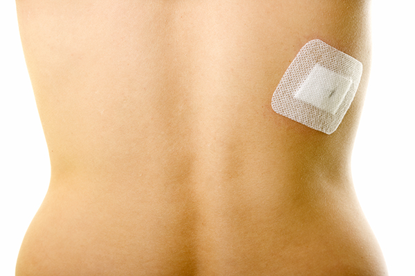
Dr. Kubeck, however, specializes in performing a minimally invasive TLIF. Dr. Kubeck first makes a small incision off the middle of your lower back and inserts a small tube through the incision until it “rests” on the spine. Using special surgical instruments, Dr. Kubeck performs the entire TLIF procedure through the tube.
By working through the small tube instead of a traditional “open” incision, Dr. Kubeck is able to significantly reduce the amount of muscle and tissue he needs to cut or disrupt, and the amount of blood loss. This typically translates to a shorter hospital stay and faster patient recovery times.
XLIF® (eXtreme Lateral Interbody Fusion)
The XLIF procedure differs from the minimally invasive TLIF in that Dr. Kubeck accesses the disc space using a surgical approach from the side (lateral) through an incision just above the hip bone rather than from the front or back.
By definition, the XLIF procedure is a minimally invasive type of spine surgery, which means that there is a smaller incision made. And a smaller incision means:
- Less muscle disruption
- Less blood loss during surgery
- Reduced operation time
- Reduced post-operative pain
- Reduced hospital stay
- Reduced recovery time

Lumbar Microdiscectomy
Dr. Kubeck performs this minimally invasive procedure to relieve pressure on the spinal nerve column caused by a herniated lumbar (lower back) disc. The goal of the procedure is to decrease pain and allow you to regain normal movement and function.
Dr. Kubeck performs the procedure using a special microscope to view the disc and nerves. Using this microscope provides a larger view, which, in turn, allows him to make a smaller incision. The smaller the incision, the less damage is made to surrounding tissue.
Symptoms
Pain and restricted movement are the most prevalent symptoms of a herniated lumbar disc. If your injury doesn’t respond to non-surgical treatments, Dr. Kubeck may suggest surgery if you:
- Have very bad leg pain, numbness, or weakness that limits your normal daily activities
- Your symptoms don’t get better after at least four weeks of non-surgical treatment
- Have new numbness, tingling, weakness, or loss of bowel/bladder control. These may constitute an emergency situation.
The Procedure
The goal of a lumbar microdiscectomy is to remove the disc material that is putting pressure on the nerves by the herniated disc. The procedure itself is performed with the patient lying face down. Dr. Kubeck then makes an incision directly over the affected disc through which he removes the damaged tissue. He then closes the small incision with sutures. Patients are typically discharged the same day or the morning after an overnight stay in the hospital.
Mobi-C® Disc Repacement
Cervical Disc Replacement
Cervical disc replacement surgery is designed to relieve the pressure on your spinal cord or nerves that may be causing pain, numbness, and weakness that can radiate from your neck to your shoulder, arm, and hand.
The benefits of cervical disc replacement surgery include:
- Maintaining normal motion in the neck
- None of the complication or issues sometimes associated with a bone graft or fusion
- A reduced chance of degenerative disc disease in adjacent cervical joints
Causes
Dr. Kubeck typically performs this surgery for patients who have a cervical disc herniation or cervical degenerative disc disease that causes chronic neck and/or arm pain that hasn’t been relieved through non-surgical treatments.
Mobi-C® Procedure
Dr. Kubeck uses the Mobi-C® artificial disc, which is designed to replicate the natural motion of the cervical spine in order to preserve range of motion in that area of the neck. Mobi-C is designed as an option to disc fusion, which limits the neck’s range of motion.
Mobi-C’s mobile core slides and rotates inside the disc, self-adjusting to the patient’s cervical spine’s movements. This means that Mobi-C can react to the normal motion in the cervical spine.

Cervical Spinal Decompression and Fusion
Dr. Kubeck performs cervical decompression surgery to relieve compression of one or more of the vertebrae located in your neck by removing structures such as bone spurs that are pressing on the nerves in your neck. Removing these structures alleviates pressure and allows the nerve to heal.
However, in some situations, there are so many bony structures pressing on the nerve that removing them may cause instability. When this occurs, Dr. Kubeck will typically perform cervical fusion to correct the instability and prevent the vertebrae from collapsing and rubbing together.
Causes
Cervical compression of the vertebrae can occur for a number of different reasons. One of the most common is the gradual wear and tear on the cervical spine that typically occurs in people over 50 years of age.
Other conditions that cause cervical compression usually develop more quickly and occur at any age. These include:
- Injury to the cervical spine
- Scoliosis (abnormal spine alignment)
- Bone disease
- Spinal tumor
- Certain bone diseases
- Rheumatoid arthritis
- Infection
Symptoms
Symptoms of cervical compression include numbness, pain, and weakness. The first line of treatment may involve non-steroidal, anti-inflammatory drugs (NSAIDs) for relief of pain and swelling, physical therapy to strengthen your neck muscles, bracing to provide support; and/or steroid injections. Except in emergency cases, surgery is typically the last resort.
The Procedure
During the fusion procedure, Dr. Kubeck inserts a spacer bone graft to fill the open disc space, which he secures in place with metal plates and screws. After the surgery, your body will begin its natural healing process and new bone cells grow around the graft. In about three to six months, the graft should join the two vertebrae and form one solid piece of bone.
